Browse through amazing photos from the world of nanotechnology and find out more about each image in the TryNano.org image gallery.
Image Gallery
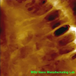
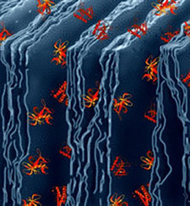
Visualization of the cell junction of human epithelial cells by AFM: High magnification image with a scan size of 7 x 7 mm2 showing the detailed structure of cell junctions. Image source: MSU Nano Manufacturing Lab
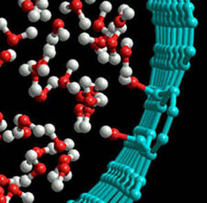
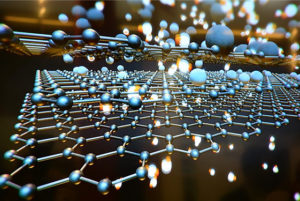
Model of water inside a carbon nanotube. Image Credit: Henry Ye, Drexel University
The tools that have allowed us to observe the previously invisible world of the nanoscale objects include special sophisticated microscopes such as the Atomic Force Microscope and the Scanning Tunneling Microscope.
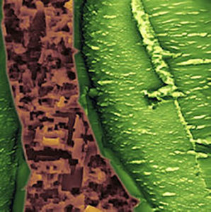
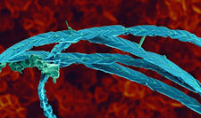
Scanning electron microscopy of wood-derived Silicon Carbide. Image Credit: Katya Vishnyakova and
Gleb Yushin, Drexel University
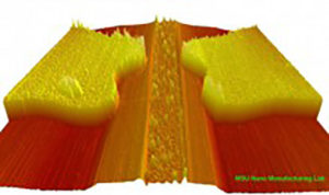
Model of mesoporous carbide derived carbon with proteins. Image Credit: Gleb Yushin, Drexel University